Retinal Disease
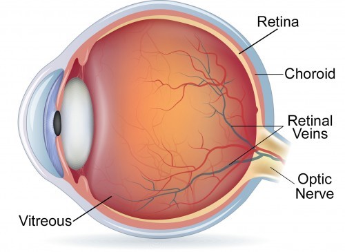
The retinal is a thin layer of light-sensitive tissue that lines the back of the eye. It turns light into electrical impulses that are sent to the brain through the optic nerve. The vitreous humor is the clear gel that fills that back of the eye.

Many retinal problems relate to the blood supply of the retina. Veins or arteries can get blocked by hardening of the arteries, embolic cholesterol plaques that break off of arteries, and inflammatory diseases. These are called retinal vein occlusions or retinal artery occlusions. Treatment may include laser treatment to prevent abnormal blood vessel growth or injections of medicine to reduce the retinal swelling caused by the blocked vessels.
Diabetic Retinopathy is a result of microvascular blood vessel damage that cause retinal bleeding, swelling and ischemia (lack of oxygen for the retinal tissue). The ischemia can lead to abnormal blood vessel growth in advanced retinopathy. Diabetic retinopathy is treated with laser treatments and injections of medications to decrease retinal swelling (edema) or reduce abnormal blood vessel growth (called neovascularization or proliferative diabetic retinopathy).
Age Related Macular Degeneration (ARMD) is an age related deterioration of the macula, the part of the retina that provides your central vision. It is usually causes a slow decrease in central vision. About 10% of patients with ARMD will develop abnormal blood vessels (this is called wet macular degeneration). Wet macular degeneration is treated by injections of medicines to decrease bleeding, swelling and new, abnormal blood vessel growth. Occasionally, laser treatment of the abnormal blood vessels is required.
Retinal holes are small holes in the peripheral retina that can lead to increased risk of retinal detachment, an ophthalmic emergency. Retinal holes are more common in nearsighted patients because the retina is thinner and more easily torn. Patients with a retinal tear or hole notice a sudden onset of many floaters in their vision and often a flashing light from the retina around the hole. If a hole or tear develops, it is usually treated with laser to decrease the risk of a retinal detachment. If the retina detaches (separates from the back of the eye), it must be repaired surgically by a retinal specialist.
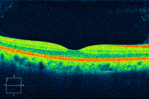
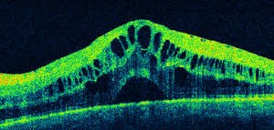
There are many more retinal disease processes not listed here. At The Eye MDs, our doctors are experts at evaluating and treating retinal problems. We have state-of-the-art equipment to evaluate the retina, such as our Ocular Coherence Tomographer that uses light waves to obtain magnified cross-sectional images of the central retina called the macula (thus the scan is called a Macular Ocular Coherence Tomography, or MOCT). MOCT scans reveal the layers of the central retina that provides our fine vision. The MOCT scan can detect swelling, retinal holes, distortion of the retina, macular degeneration and other retinal pathology (see MOCT image below).


